View Video
Age-related macular degeneration (AMD) is one of the most common causes of poor vision after age 60. AMD is a deterioration or breakdown of the macula. The macula is a small area at the center of the retina – the light-sensitive tissue lining the back of the eye. The macula allows us to see fine details clearly and perform activities such as reading and driving. The macula makes up only a small portion of the retina, but it is much more sensitive to detail than the rest of the retina, called the peripheral retina. The peripheral retina is responsible for side vision. Damage to the macula can dramatically affect your vision while damage to the peripheral retina is usually well tolerated with only minimal affect on functional vision. In fact, AMD is responsible for 90 percent of new cases of legal blindness in the United States. The visual symptoms of AMD involve loss of central vision. They include blurriness, dark areas or distortion in your central vision, and perhaps permanent loss of your central vision.
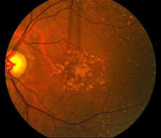
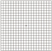
90 percent of people who develop macular degeneration have the dry form. It is characterized by aging and thinning of the tissues of the macula. Tiny yellow or white deposits of fatty protein called drusen accumulate under the retina. With dry AMD, vision loss is usually gradual. People with this disease need to monitor their vision closely. Any changes in vision need to be reported to the ophthalmologist right away. A simple, at home test, called an Amsler grid can be used to help monitor vision.
About 10 percent of those with AMD have the wet form. It is characterized by sudden, severe loss of central vision. Typically, damage to the layer under the retina allows abnormal blood vessels to grow into the space between the retina and underlying tissues. These blood vessels leak fluid or blood causing significant distortion to the vision and eventually permanent scarring of the macula.
Genetic changes appear to be responsible for approximately half the reasons an individual develops macular degeneration. Additionally, there are other risk factors for developing the disease:
- Part of the body’s natural aging process
- Heredity – if you have a close family member with the disease
- Smoking
- High blood pressure
- Abnormal cholesterol levels
To check for AMD, your eye doctor must dilate, or widen, your pupils using eye drops to gain better visualization of your retina. This will allow for a much clearer evaluation of your retina using an ophthalmoscope, a device used to see fine details of the retina. Specialized tests can be performed to aid in the diagnosis and characterization of AMD. Optical coherence tomography (OCT) is a machine that takes special pictures of your retina helping to detect very subtle amounts of fluid and abnormal tissue. Also, fluorescein angiography may be performed. During this test, a dye called fluorescein is injected into a vein in your hand or arm. Photographs of your retina are taken in succession that allows the doctor to follow the flow of that dye through the vessels of your retina looking for abnormal blood vessels and fluid leakage. These vessels and fluid are very difficult to see without this test. The results of these tests help guide treatment.
At this time, there is no single proven treatment for the dry form of macular degeneration. However, a large scientific study did show that certain antioxidant vitamins and zinc may slow the progression of the disease by about 20 percent. This study was called the Age-Related Eye Disease Study (AREDS). The formulation of vitamins used in the study were as follows:
- Vitamin C – 500 mg
- Vitamin E – 400 IU
- Beta carotene – 15 mg
- Zinc oxide – 80 mg
- Copper – 2 mg
Another study is currently underway to evaluate the benefits of lutein and fish oil (omega-3). Studies have shown that eating dark leafy greens, and yellow, orange, and other colorful fruits and vegetables, rich in lutein and zeaxanthin, may reduce your risk for developing macular degeneration.
Researchers have found that a chemical called vascular endothelial growth factor, or VEGF, is critical in causing abnormal blood vessels to grow under the retina. Scientists have developed several new drugs that can block the trouble-causing VEGF known as “anti-VEGF” drugs. They help block abnormal blood vessels, slow their leakage, and help reduce vision loss.
Treating the wet form of AMD may involve the use of anti-VEGF treatment, thermal laser treatment, or photodynamic therapy (PDT). Treatment typically reduces but does not eliminate the risk of severe vision loss. Anti-VEGF medications are a step forward in the treatment of wet AMD because they target the underlying cause of abnormal blood vessel growth. This treatment offers new hope to those affected with wet AMD. Although not every patient benefits from anti-VEGF treatment, a large majority of patients achieve stabilized vision, and a significant percentage can improve to some degree. This medicine is administered directly into the eye during an in office procedure. This procedure usually needs to be repeated several times at 6 to 12 week intervals until the disease process is stabilized. The other two forms of treatment involve the use of lasers to treat the new, leaky blood vessels directly. They are typically used as adjunctive treatment along with anti-VEGF treatments. Patients who have suffered from the wet form of AMD for months to years will likely not have significant benefit from these treatments and may not be offered therapy. These treatments work best on newly diagnosed wet macular degeneration.